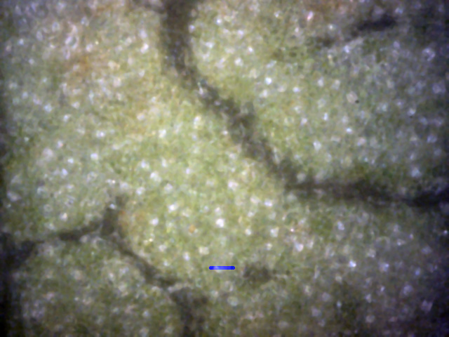
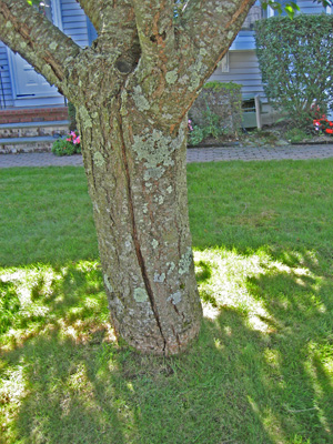
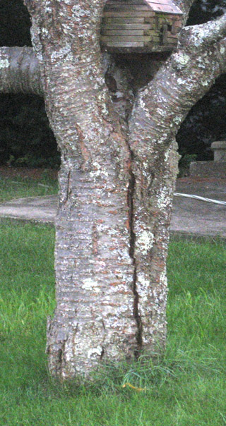
After seeing so many diseased leaves, I decided to check out an apparently disease-free Kwanzan Cherry.
It was about 12' high and appeared to be about 10 years old.
There were no apparent bark or leaf problems.
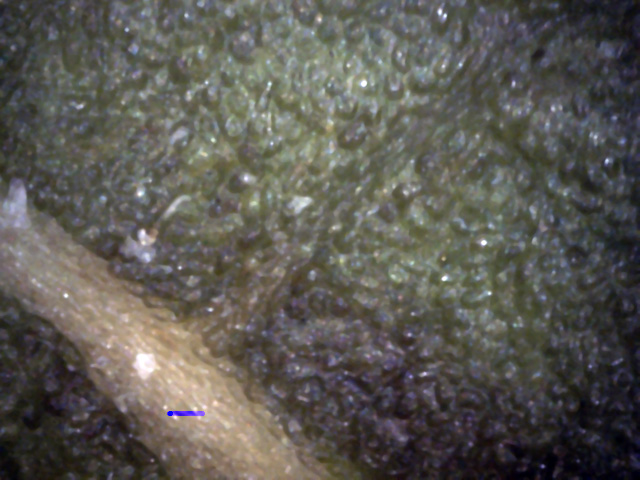
The pictures below were taken in early August, using a 400x microscope.
The blue lines are 50 micrometer scale bars. This is about the diameter of a white canker spore.
1
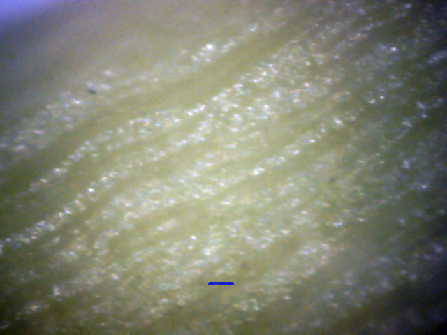
This leaf top, near the vicinity of a stem, is virtually blemish-free.
There is only the slightest evidence of a spore.
The remainder of the leaf had no evidence of any disease.
2
Leaf stems can be badly infected, but, as this picture shows, this leaf stem
is in excellent shape, with no signs of disease.
4
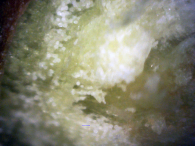
The first hint of a problem came when I ripped a leaf off of a branch and examined the leaf scar.
Here you can see what appear to be very young spores just inside the leaf's green cortex layer.
While very hard to see, they appear to have that characteristic cleft in them.
5
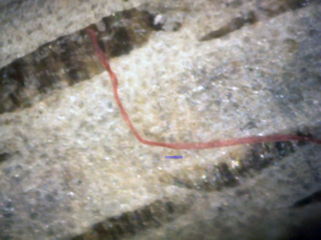
While the bark of a twig infected with white canker will often be
in poor shape with lots of spores, this twig's bark was unusually clean.
I had to look hard to find the single infectious ribbon shown here
(note that it appears to be emerging from a hole in the bark).
Strangely, the color of the ribbon is pink.
This ribbon went on for quite a bit and then entered the wood again.
6
←
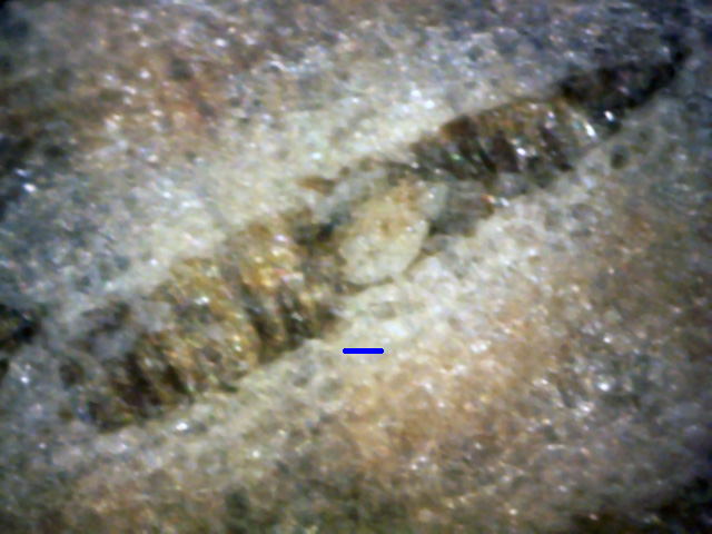
This might be a spore emerging from the wood (red arrow). Note that it seems
to emerge at a bark split.
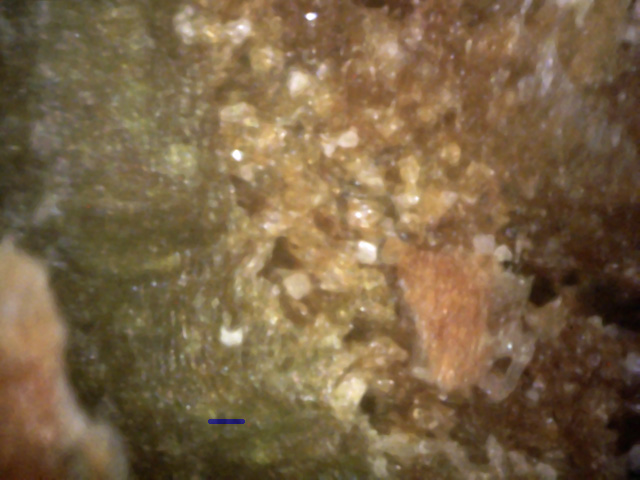
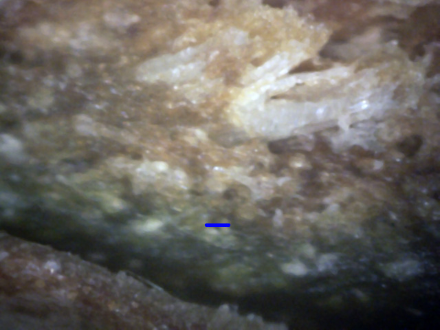
While the twig and bark pictures seem to show no white canker infection, the final "say" is usually
determined by twig cross-section views, as shown in the following 400x microscope views taken
of the cross-section of a 3/16 inch diameter twig. The bark is on the lower-left of each picture.
7
←
The green cortex has no voids nor growths inside it, and has no gaps between it and the outer bark.
However, the phloem tissue to the right contains some gaps which seem to host white hyphae.
A red arrow shows the end of one such hypha, which is growing within a void it probably has eaten out
as it was growing along the branch.
8
↑
↑
This view more clearly shows evidence of a white canker infection.
The green cortex is thin, contains white canker growths, and has separated from the bark.
Furthermore, the adjacent phloem has several large clumps of white canker hyphae (red arrows).
In summary, this apparently healthy tree shows virtually no evidence of white canker when examining the
leaf or twig surfaces. That correlates with the general appearance of the tree.
Twig cross-sections, however, seem to tell a different story, since they expose the phloem and cortex -
the focus of white canker. Here we see the very beginnings of a white canker infection.
Consequently, we can expect this small tree to begin a steady decline in a year or two as the white
canker slowly chokes off its nutrient supply.
